- History Home
- People, Leadership & Service
- A Legacy of Excellence
- History & Impact
- Meetings Through the Years
- Resources
Memoir - Alexander WlodawerMemoir | Publications | Curriculum Vitae | Videos | Slides | Articles | Obituary ACA Living History2019
I decided to become a scientist rather early on—at the ripe age of four. My mother was a biochemist at the Nencki Institute of Experimental Biology in Warsaw, Poland. One day I offered to Professor Włodzimierz Niemierko, then director of the Institute, my future services subject to successful graduation from kindergarten, school, and university. Little did I know that I would actually accomplish that goal, but, to tell the truth, any thoughts about crystallography were definitely not on my mind then. Moreover, although those promising beginnings took place in Poland, almost my entire scientific career has been connected with the United States, a fact that I attribute to several circumstances.
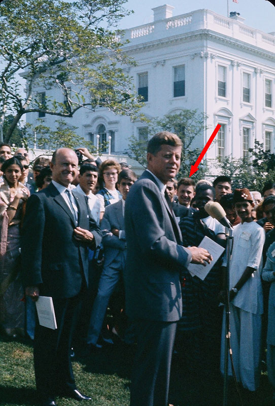
A visit to the White House in 1962, hosted by President John F. Kennedy. I am identified by the arrow.
When I was in high school, I was selected to become a member of the Polish delegation attending a meeting of the American Junior Red Cross on the hundredth anniversary of the establishment of the Red Cross in the United States. Luck was with me: English was not taught in my school, but my parents insisted early on that I study the language, so I did not have much competition in the selection process. That meeting in the summer of 1962 truly changed not only my own life but also the lives of many other participants. A visit to the White House hosted by President John F. Kennedy (Fig. 1) was truly inspiring, and a visit to the United Nations headquarters in New York provided an impetus for another young meeting participant, Ban Ki-moon, to become a diplomat and later occupy the most important office in that same building for a decade. My own goal was also set: I would finish my high school and university studies and go to the United States for graduate studies.
When the time came to choose the direction of my university studies, I abandoned the idea of life sciences and decided to study physics. I followed that track for three years until 1966, when I had to select my specialization. That same year, Professor David Shugar started a completely new program in biophysics in Warsaw. I joined its very first class and decided to work on my master’s thesis at none other than the Nencki Institute. However, rather than conducting experiments in physics, I dabbled in neurophysiology of vision but quickly convinced myself that torturing cats should definitely not become my future career.
At that time, many young people in Poland became very active politically and secret groups were discussing how to improve socialism to the point that it would actually deliver on its promises. Major political upheavals took place during and after March 1968, which strongly encouraged me to emigrate. Although I was already accepted to a graduate program in neurobiology at the University of Iowa, I also applied for doctoral studies at two universities in California, UCLA and Caltech. The major impetus was geography: I was a mountain climber, and realized in time that there were no mountains in Iowa. I later discovered that my application to Caltech was never considered, since I had been unable to send them the $10 application fee. However, I was not only accepted to the Molecular Biology program at UCLA but also awarded a stipend. Thus, it was not Iowa, but California, and not neurobiology, but molecular biology. The only problem was how to get there.
Due to some rather obscure American regulations, I could apply for a refugee U.S. visa only in Italy. Therefore, I went to Rome, filed visa applications, and waited. In a serendipitous development, I was hired as a completely unqualified technician in a laboratory in the Istituto Superiore di Sanita. The head of the laboratory was Rita Levi-Montalcini, who some years later became a Nobel laureate, and the area of study was a small protein called nerve growth factor (NGF). I became completely fascinated by this hormone that directs the growth of neurons. Of course, it was not at all clear to me that this would become important much later.
I came to Los Angeles in the summer of 1969 and started my graduate studies the day after my arrival. That same year, a young scientist named David Eisenberg moved from Caltech to UCLA to become an assistant professor. I became one of his first graduate students. David decided to establish at UCLA a new area of investigation, namely protein crystallography. It was barely a decade since the first protein structures had been determined by Max Perutz and John Kendrew, and only a few places in the world were engaged in such studies. I certainly did not plan on becoming a crystallographer when I started my graduate work, but I was very quickly converted and realized that this should be the field of my specialization.
For the next four years, I tried to solve the crystal structure of rabbit muscle aldolase. I cannot say that the work was going well, and there was no structure by the time I was ready to write my thesis. However, it was still possible at that time to graduate without solving a protein crystal structure and by publishing only a single paper—thus, Ronald Reagan’s signature was finally placed on my Ph.D. diploma (he was then the governor of California). While at UCLA, I tried to interest David in NGF, but he did not bite.
My next move was to look for a postdoctoral position. I was offered an opportunity to work with Martha Ludwig in Michigan, but since I knew there were no mountains worth climbing in that state, I declined. On the other hand, Brian Matthews at the University of Oregon must have learned that my Ph.D. thesis presented little experimental data, so he very politely turned me down. My luck somehow prevailed, however: I contacted Eric Shooter, a professor at Stanford University and one of the major players in the NGF field. Eric became interested and promised to support my quest for the structure of this protein, but since he did not have funds to support me, he made a deal with Keith Hodgson, who at that time was starting a project to utilize synchrotron radiation as a source of X-rays for protein crystallography. Thus, I could work on both methods development and structure determination.
The summer of 1974 was the most successful period in my career as an experimental crystallographer. I crystallized not only NGF but also two other proteins, L-asparaginase and monellin. At that time, just crystallization of a protein alone was sufficient for a full publication, even in Proceedings of the National Academy of Sciences. However, the development of a synchrotron beamline as a source of X-rays was a much slower project, and I did not have any equipment to collect X-ray diffraction data at Stanford. I ended up flying regularly to Oregon, so Brian Matthews was stuck with me despite his earlier decision.
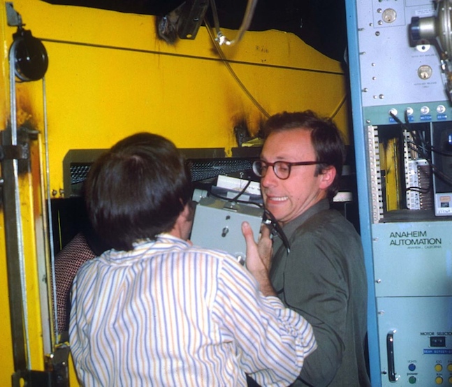
Keith Hodgson (with his back turned) and I installing a precession camera in the hutch at the
Nevertheless, my main project at Stanford was the development of the first synchrotron beamline for protein crystallography. That work was directed by Keith Hodgson, with further participation by Margueritte Yevitz Bernheim and a graduate student, the late James Phillips. We were joined by Julia Goodfellow (now Dame Julia) a year later. To say that our facilities were primitive is to overestimate the true state of affairs. Our only detector was an Enraf-Nonius precession camera (Fig. 2) that could be used with Polaroid films for alignment or with multiple packs of radiology films for “data collection.” The latter films could be later scanned to provide some numerical data, but usually we just looked at them and extrapolated the speed of data collection to the future when everything would work perfectly (it never did at that time).
As is often the case in protein crystallography experiments, we started with crystals of hen egg-white lysozyme, which are easy to grow and diffract X rays very well. We were quite happy when a precession photograph could be obtained in as little as two hours—that was actually the duration of a single fill of the synchrotron storage ring. We later used this beamline to collect diffraction data for proteins such as NGF, L-asparaginase, azurin, and rubredoxin. Most crystals of these proteins were too small to provide measurable diffraction with standard laboratory X-ray tubes, so we considered the use of synchrotron radiation to be quite successful. Experiments involving rubredoxin, performed with Lyle Jensen and his colleagues at the University of Washington were particularly important, since we tuned the wavelength to match the absorption edge of iron, thus maximizing the anomalous signal. We were quite pleased to see even by the naked eye that there were differences between the intensities of Friedel mates (the central projection in the space group R3 is non-centrosymmetric). Those very early experiments clearly proved that the tunability and high intensity of the synchrotron X-ray beam would ultimately revolutionize protein crystallography.
Running experiments was exhausting, since the beam was dumped every two hours and it was necessary to adjust the camera after every fill. My longest single experiment took six nights and five days, with sleep possible in—at most—two-hour increments (on the floor, under a table). We felt pressure to get some positive results before others would beat us to it and, by mid-1976, we finally published our preliminary results in Proceedings of the National Academy of Sciences—just in time, since the results from Deutsches Elektronen-Synchrotron (DESY) in Hamburg came out soon thereafter, and another group in Novosibirsk was also developing a protein crystallography beamline.
My next move—in 1976, to the National Bureau of Standards (NBS, now the National Institute of Standards and Technology) in Maryland—was a direct result of meeting Hal Wyckoff, a well-known crystallographer from Yale University. Hal and I had many previous conversations about building crystallographic instruments, so, without my knowledge, he recommended me to David Davies as a suitable candidate to develop a neutron diffraction facility. Considering that the only trained American macromolecular neutron crystallographer had just quit that job, I did not have many competitors, was selected, and moved to Gaithersburg, Maryland.
While working at the NBS, I tried to balance two requirements: developing a neutron diffraction station that utilized a completely unique detector (that was what I was paid for), and continuing my work on the structures of NGF and L-asparaginase (in my “free” time). Fortunately, David Davies assigned to me a corner in his laboratory in the attic of Building 2 on the Bethesda campus of the National Institutes of Health (NIH), and there I struggled with structure determination, without much apparent success. On the other hand, my main project, which involved construction and utilization of a neutron diffractometer capable of measuring data from protein crystals, progressed much better. To a large extent, that progress was due to two colleagues with very extensive knowledge of neutron technology, Antonio Santoro and Ted Prince. They came up with the idea of building a flat-cone diffractometer utilizing a 1-meter-long linear detector and helped me with writing the operating software. I was later joined by my first postdoctoral fellow, Lennart Sjölin, who very successfully continued the process of software development. My predecessor, John Norvell, had already grown crystals of ribonuclease A (RNase A), the largest having a volume of 100 mm3; thus, the course of action for the next eight years was set.
Lennart and I initially concentrated on the determination of the crystal structure of RNase A based on neutron data alone, but we quickly realized that this might not be the best way of proceeding. However, discussions with Wayne Hendrickson, with whom I would meet quite regularly during the Washington Crystal Colloquia—organized by no less than a future Nobel laureate, Jerome Karle—led us to adapt Konnert and Hendrickson's program PROLSQ for joint X-ray and neutron refinement. This approach allowed us to publish quite significant data on the protonation states of residues such as histidine and on amide hydrogen exchange.
Subsequently, we decided to investigate another small protein, bovine pancreatic trypsin inhibitor (BPTI). In retrospect, our BPTI work was much more important than our RNase A work, as BPTI became a prototype for the development of macromolecular NMR and for computational methods interpreting the folding, structure, and dynamics of proteins. Since BPTI was originally studied in Munich, I established a very fruitful collaboration with the future Nobel laureates Robert Huber and Hans Deisenhofer. X-ray data personally collected by Robert, merged with our neutron data, were used for joint refinement, leading to the first truly atomic-resolution (1 Å) protein structure to be deposited in the Protein Data Bank (PDB). The structure of the even-smaller protein crambin was refined earlier but deposited later by Martha Teeter. The BPTI project also painfully taught me what happens if diffraction data are not deposited together with the coordinates: some years later, neither Robert nor I could find the diffraction intensities, which became permanently lost.
An important event happened a few years after my move to the NBS. The 1978 Congress of the International Union of Crystallography was going to take place in Warsaw, so I decided to make an attempt to attend. I was by then both a U.S. citizen and a civil servant, so I thought that maybe I had a chance to obtain a visa. Indeed, my visa materialized a few days before the meeting, largely due to the efforts of Jerome Karle (who was later banned from visiting Poland as a punishment for forcing the government to issue visas to undesirable elements, such as myself). While in Warsaw, I met Tom Blundell, one of the top British crystallographers of the second generation. Tom and I discussed the NGF stalemate in considerable detail and came to an understanding: his laboratory would take over the project, but I would be kept in a supporting role. That agreement held for the next 13 years—that was how long it took to finally determine the structure of this very small protein. The results were worth it, though: the structure, published in Nature in 1991, elucidated a newly discovered fold that included a cystine knot, later found in many other important proteins. Tom held his part of the bargain and I was included as a co-author of that paper, even though by then we were more competitors than collaborators. Another bonus of that Congress was meeting Liang Dong-cai from China, who provided me with a few large crystals of insulin that we used in a subsequent neutron diffraction study.
I continued my neutron diffraction work through 1984, when I was asked by the NBS management to de-emphasize the neutron diffraction work and to establish a new laboratory devoted primarily to protein X-ray crystallography. I recruited my former postdoctoral fellow Maria Miller, as well as Gary Gilliland, and we started several new projects. Gary inspired my interest in the structure of chymosin, a typical aspartic protease from the pepsin family. I will never forget a talk that I gave at Birkbeck College in London, during which Tom Blundell, looking at a slide showing all 2500+ atoms of this enzyme, asked me why Tyr75 was in the wrong place. It turned out that it was not, but I clearly did not fully understand the implications of its movement.
Another important event in my career that could be traced to attending a scientific meeting took place in 1986. I participated in a Congress of the European Crystallographic Association in Wroclaw, Poland (by then nobody cared if I would come to Poland or not). The meeting was very sparsely attended by scientists from the West, with a notable exception of the future Nobel laureate Ada Yonath, who caused the organizers a major headache with her request for help in procuring enough gasoline so that she could drive her car back to Berlin. However, at this Congress I met a distinguished Polish crystallographer, Professor Zofia Kosturkiewicz, who suggested that I accept her former student Mariusz Jaskólski as a visitor to my laboratory. Indeed, Mariusz came to the U.S. a year later, and we then started our very successful collaboration that continues until today and that has resulted so far in more than 40 joint publications.
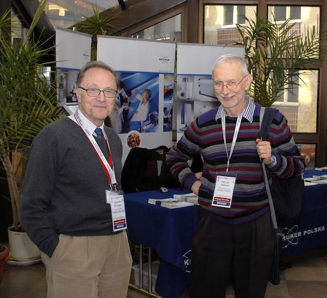
Mariusz Jaskólski and I attending a Multi-Pole conference in Warsaw, Poland, in 2011.
In the meantime, the laboratory at the NBS was expanded by the addition of a husband-and-wife team, Robert Harrison and Irene Weber—the former as my postdoctoral fellow, and the latter as an independent scientist. The range of projects was increased, but the facilities were rather limited, especially since we were largely separated from the biological community. I felt that it was time to move on.
My move to the National Cancer Institute (NCI) was an indirect result of Joel Sussman’s sabbatical at the NIH. Joel worked closely with us on structural investigations of DNA duplexes containing unpaired bases and spent lots of time in our laboratory. In 1986, he told me about a plan to start a structural biology laboratory at the NCI in Frederick, Maryland, and encouraged me to apply for a group leader position (he himself applied for the position of a lab chief). However, Joel ultimately decided to accept the position of the director of the PDB (then at Brookhaven), and George Vande Woude, the head of the Frederick program, offered me the lab chief position. I accepted and moved to Frederick in 1987, with Irene Weber assuming a group leader position, and with Ron Rubin joining us as a group leader a little later.
The moment of transition between the NBS and NCI happened when my laboratory became engaged in a new and exciting research area, namely structural investigations of retroviral proteases (PRs). As is often the case, the start of the project was quite fortuitous—through my introduction to Jonathan Leis, who at that time worked at the Case Western Reserve University in Cleveland, Ohio. Jonathan had been working for a long time on biochemical characterization of various retroviral proteins and had successfully purified milligram quantities of PR from Rous sarcoma virus (RSV, now usually called avian sarcoma virus, or ASV). We immediately decided to investigate its three-dimensional structure as a stand-in for the structure of the much more medically important enzyme encoded by human immunodeficiency virus type 1 (HIV-1).
One should remember that the 1980s were the time of a looming global epidemic caused by an unknown virus that was triggering an invariably fatal disease called acquired immunodeficiency syndrome (AIDS). This virus was considered to be the most dangerous emerging threat to human health. HIV-1 was conclusively shown to be the causative agent of AIDS in 1983, and its RNA genomic sequence was first published in 1985. At that time, the HIV-1 genome was shown to encode only three enzymes, one of which is PR. This protein plays a crucial role in the life cycle of HIV-1 and of other related retroviruses, such as RSV, since it is needed to cleave the precursor viral polyproteins into mature, individual proteins. Inactivation of PR was shown to prevent viral particles from maturing into their infective form, thus making PR a potential target for antiviral drugs. However, genuine proteins from HIV-1 were very difficult to come by at that time, although, as we found out later, researchers at the Merck Sharp & Dohme laboratories had succeeded in large-scale production and purification of recombinant HIV-1 PR.
A hypothetical model of HIV-1 PR was built in 1987 in a bold modeling exercise performed in England by Laurence Pearl and William Taylor, despite almost no sequence conservation between cell-derived proteases and retroviral PRs. We were familiar with that model, looked at it with interest, but did not fully believe in its correctness. Besides, that was HIV-1 PR, and the enzyme in our hands was RSV PR, which had only ~30% sequence identity with HIV-1 PR.
Although we entered the race to determine the structures of retroviral PRs relatively late, a series of serendipitous events contributed to our success. Crystals of RSV PR were grown by Maria Miller within a month of receiving the protein. Derivatization of the crystals with a uranyl compound, an excellent anomalous scatterer of CuKα radiation, yielded a single-site derivative (which marked, as it later turned out, the active site) that enabled the proper choice of the space group enantiomorph and helped in setting some additional derivatives in common origin and handedness. J. K. Mohana Rao and Mariusz Jaskólski were crucial participants in that phase of the project. The electron density map, based on multiple isomorphous replacement phases from the four best derivatives, allowed us to trace the main chain of the dimeric molecule, and the atomic model of RSV PR was complete in October 1988.
As soon as the first RSV PR model was complete, Irene Weber built a homology model of the HIV-1 enzyme. The model looked very plausible: it had all the features of the template, with differences limited to the loop regions. The structure of RSV PR was published in Nature in early February 1989. A week later, in the same journal, the crystal structure of HIV-1 PR was unveiled by Manuel Navia, Paula Fitzgerald, and co-workers from Merck Sharp & Dohme, and that same week, Irene’s model was published in Science. After the first burst of joy, suddenly there was consternation because the crystal structures of the RSV and HIV-1 PRs, while similar in the basic features, also showed some perplexing differences, especially in the C-terminal region of the molecules. Whereas the RSV PR model had a clear α-helix, the HIV-1 PR structure had a straight β-strand, and the topology of the dimer interface was completely different. Instead of the interlaced termini with three inter-subunit β-sheet connections found in the RSV PR, the HIV-1 PR crystal structure had a hairpin with only one area of inter-subunit contact, and a disordered N terminus. The latter difference was not trivial; rather, it had profound consequences for the dimer stability and for the PR’s ability to excise itself from the viral Gag-Pol fusion polyprotein synthesized in the infected cell. Moreover, the question about the correct features of retroviral PR was not purely academic, because an accurate HIV-1 PR model was badly needed for a structure-guided design of inhibitors that might be developed into AIDS drugs.
The situation in February 1989 became rather uncomfortable: which HIV-1 PR model should be used for designing AIDS drugs? Which one was correct? The dilemma could be resolved only by experiment, but the main question was how to obtain the protein. Help came from Stephen Kent, then at the Caltech, who was pioneering the methodology of protein synthesis using a purely chemical process. He and Jens Schneider quickly sent us 0.2 mg of chemically synthesized HIV-1 PR, enough to grow a few crystals. Our molecular replacement calculations had to rely on Irene’s model of HIV-1 PR, as the coordinates of the Merck structure were not made available. However, more material was needed to produce heavy-atom derivatives, because it was critical to obtain phase information experimentally, to avoid model bias, and to produce an independent model of the protein. More protein was also needed for cocrystallization trials with inhibitors. The Kent group set a precedent by producing for us, within a period of just two weeks, milligram quantities of HPLC-purified enzyme for successful crystallographic studies. The definitive structure of the HIV-1 PR apoenzyme, showing its agreement with the RSV PR-derived model, was published in Science in August 1989.
The next goal was to determine the structure of HIV-1 PR in complex with inhibitors. The first such inhibitor, MVT-101, was provided to us by Garland Marshall (Washington University). Cocrystals with the synthetic enzyme grew overnight, and we were able to complete and publish the structure of the complex four months after the publication of the structure of the apoenzyme. It is worth stressing that the coordinates of the synthetic HIV-1 PR:MVT-101 complex were deposited in the PDB in April 1990 and, for the two most critical years, were the only ones freely available to all researchers worldwide who were working on the design of specific retroviral PR inhibitors, although we were not directly involved in such efforts.
Some PR inhibitors were developed into potent drugs for treating HIV-1 infection. The first HIV-1 PR inhibitor to become a drug, Saquinavir (Ro-8959), was developed by Roche and approved for clinical use in December 1995—only six years after the structure of the first inhibitor complex had been published and less than seven years from the moment an experimental model of the protein saw the light of day.
With very significant participation by Alla Gustchina, who had studied the structure of pepsin in Moscow and was thus well acquainted with aspartic proteases, we continued the project by investigating retroviral PRs from other sources, such as FIV, EIAV, XTLV, and XMRV. Our focus was on models of potential resistance to anti-HIV drugs, as well as on the function of these enzymes in carcinogenic viruses. These crystallographic studies allowed us to establish some general principles of the relationship between structure and activity of this family of enzymes. The work on HIV-1 PR was later continued by Irene Weber after her departure from the NCI, first to Philadelphia, and later to Atlanta. It is generally recognized that determining the structure of HIV-1 PR has been the springboard for the development of successful rational drug design strategies not only in that particular case but also for other pharmacological targets, taking the idea from a flimsy dream to practical reality.
Our involvement in AIDS-related projects did not end with structural studies of retroviral PRs; a few years later, we selected as our next target the retroviral integrase (IN), which is the enzyme responsible for inserting a DNA copy of the viral genome into the host genome. That project was started in a manner similar to what was previously done for PR, namely by using RSV IN as a stand-in for HIV-1 IN, which was not available to us at that time. This was again a close collaboration not only with Jonathan Leis but also with Anna Marie Skalka from the Fox Chase Cancer Center. The central person working on this project was Grzegorz Bujacz, who came to my laboratory in 1993, but Mariusz Jaskólski was very much involved in this work as well, even though by then he was spending almost all of his time in his own laboratory in Poznan, Poland. Whereas we determined the structure of the catalytic domain of RSV IN a few months after the structure of the corresponding domain of HIV-1 IN was determined by Fred Dyda and David Davies at the NIH, the detailed picture of the active site, especially the mode of binding of the key divalent metal cations, was much clearer in our model. Crystals of RSV IN were so good that another postdoctoral fellow, Jacek Lubkowski, even managed to extend the resolution to a truly atomic level.
Another area of research in my laboratory that was largely AIDS related was the result of a collaboration with the NCI group led by Barry O’Keefe. Barry led an effort to identify natural products with anticancer or anti-HIV properties, and promising hits came from identification of several lectins (carbohydrate-binding proteins) capable of preventing transmission of HIV-1 from the cells of the host to the cells of the recipient. The mode of action of lectins involves interference with the glycan shield on the surface of HIV-1: by specifically interacting with the carbohydrates that cover surface glycoproteins of the virus, lectins prevent its attachment to cell-surface receptors. We investigated the structure and activity of several such lectins, including cyanovirin, scytovirin, and griffithsin. Our work on cyanovirin, conducted together with Marius Clore and Angela Gronenborn (then both at NIH), has assisted in characterizing this lectin as a potential agent for preventing HIV-1 infection (it is now in clinical trials). Our structural studies of griffithsin led to re-engineering this protein with the aim of improving its antiviral properties. This lectin is also in current clinical trials as a topical anti-HIV agent.
While the AIDS-related projects were diligently pursued in my laboratory, I did not forget about my old love, namely L-asparaginase. That enzyme is still a successful anticancer drug, especially for acute childhood leukemia. Although our L-asparaginase project was moving very slowly, by 1988 we published a first glimpse of the structure of the enzyme from Acinetobacter glutaminasificans at a rather low resolution of 3 Å. Whereas the general shape of the molecule was correct, the chain tracing was a good example of liberal application of imagination, with almost two-thirds of it traced backwards. It took another five years until that very deficient model helped Amy Swain to solve the structure of the E. coli enzyme, and that time we got it right. Mariusz Jaskólski and Dominique Housset, a postdoctoral fellow from France, were very much involved in that project as well. Mariusz later continued in his lab in Poznan structural and enzymatic studies of plant asparaginases, enzymes with similar activity but completely different fold. We subsequently managed to obtain a truly atomic resolution structure of the enzyme from Erwinia chrysanthemi, at that time (2003) the largest such structure in the PDB. At that stage the project went dormant, but it was resuscitated in our laboratory in 2017 by Jacek Lubkowski, with the aim to finally determine the exact mechanism of catalysis by this unique enzyme.
How did I become involved in looking at proteins at atomic resolution? Of course, as with most of my scientific career, this happened through serendipity. In the mid-1990s, Fred Dyda and I realized that more than a dozen crystallographers working at various institutes of the NIH could make very good use of our own synchrotron facility. We managed to obtain support—both moral and, more importantly, financial—from the top management of the NIH and secured access to beamline X9B at the Brookhaven synchrotron. However, having access to the beamline was clearly not enough; we needed someone who could professionally operate it. Here, luck was with us: Zbyszek Dauter, who for almost 10 years had worked at the EMBL DORIS beamlines in DESY Hamburg, was persuaded in 1997 to move to Brookhaven. This was a significant loss for the European crystallographic community but a clear win for us, since Zbyszek has many talents and has been known for years as not only a great crystallographer but also a superb collaborator. Since he excels at working at the resolution of 1 Å and beyond, engaging him in projects that involved studies at atomic resolution benefited us tremendously. Many such projects have been completed through our collaboration during the last 20 years.
With our extensive involvement in structural studies of proteases, it is not surprising that my laboratory has also been active in investigating their inhibitors. Indeed, as mentioned earlier, I had been working on the structure of BPTI while at the NBS. Much more recently, our colleague from Brazil, Maria Luiza Oliva, provided us with a number of plant-derived inhibitors of enzymes from the trypsin family. These small proteins appear to exhibit anticancer properties; thus, knowing their structures and the mode of interactions with their targets could be quite important. Indeed, we determined crystal structures of a number of these inhibitors, including a protein named CrataBL, which, in addition to being a protease inhibitor, is also a lectin, thus tying together different threads of work in my laboratory.
Another major area of research in my laboratory began after Alla Gustchina returned from a brief trip to Moscow with purified samples of two cytokines, IL-3 and IL-4. Whereas we never made any progress with IL-3, the structure of IL-4 was determined by Alla and Sasha Pavlovsky and was published in 1992. Structural investigations of cytokines and growth factors were very topical at that time, and it is not surprising that as many as four structures of IL-4 were published almost simultaneously, two determined by crystallography and two by NMR. A comparison of these structures, published jointly by all four groups, provided an interesting baseline for establishing the relative advantages and disadvantages of the two methods of structure determination. Our interest in IL-4 led us to further work on IL-10 (primarily by the late Sasha Zdanov), IL-8, IL-19, IL-24, and IL-28 (IFN-λ), as well as on complexes of cytokines and JAK kinases with the extracellular and intracellular fragments of their receptors, respectively. This project is still actively pursued, principally by Jacek Lubkowski.
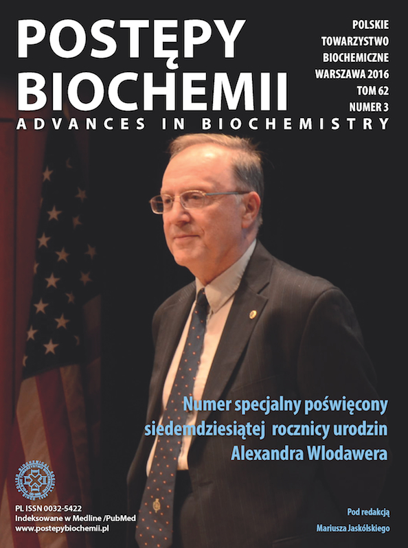
It is not a coincidence that many names mentioned in this memoir are Polish. Although I never explicitly tried to find collaborators in that country, my contacts in Poland resulted in several scientists visiting my laboratory, and some staying for many years. Additionally, many distinguished crystallographers with Polish roots are very active and successful around the world. Thus, the “Polish Crystallographic Mafia” came into existence—in addition to my colleagues mentioned above, it also includes many others. Wladek Minor, well known as a co-author of the HKL3000 package, has been particularly involved in our efforts to maintain and enhance the quality standards of macromolecular structures deposited in the PDB. Years ago, I was involved in the first crusade to make deposition of atomic coordinates of published structures mandatory—it is hard to believe today that, at that time, many prominent protein crystallographers were fervidly opposed to such a policy. However, it became the law of the land, followed some years later by the requirement to deposit experimental structure factors as well. In the last few years, we have become, together with some other colleagues who do not claim any Polish roots, self-appointed policemen monitoring the PDB, plucking rotten apples and rectifying less-severe errors of selected structures. I think that these efforts may ultimately turn out to be quite important, since the presence of bad apples in the PDB bushel is guaranteed to cause serious problems in meta-analyses, in particular by biasing projects that might lead to the creation of new drugs. Other efforts of the Mafia included the organization of meetings entitled Multi-Pole Approach to Structural Science, as well as editing the latest textbook of protein crystallography. One indication of our success was that Mariusz Jaskólski and I received in 2015 the first-ever Polish-American Scientific Collaboration Award given by the Foundation for Polish Science and the American Association for the Advancement of Science. We were very proud of being selected in a highly competitive contest encompassing all fields of science. My photo taken during the award ceremony was used for the cover of a special edition of the Polish journal Postępy Biochemii (Advances in Biochemistry), for an issue celebrating my 70th birthday.
A cover of the special issue of Postępy Biochemii (Advances in Biochemistry).
I would like to think that being invited to write this memoir does not indicate that my scientific life is over—I certainly hope this is not the case, since quite a few projects are still a long way from being completed. I have been blessed with having excellent mentors, with being able to work in well-equipped laboratories, and, most importantly, with having superb collaborators, who were principally responsible for whatever successes my laboratory could claim. I am very grateful to all those already identified, and to the many individuals whose names I did not have a chance to mention. Thank you all! |