- History Home
- People, Leadership & Service
- A Legacy of Excellence
- History & Impact
- Meetings Through the Years
- Resources
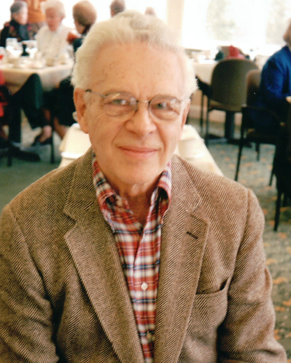
Memoir - David Sayre (1924 - 2012)Memoir | Publications | Curriculum Vitae | Videos | Slides | Articles | Obituary ACA Living HistoryDavid Sayre2010
Q. What are the main contributions that you have made? There are two. Much the more important is the concept of X-ray diffraction microscopy, dating from Sayre (1980). I was then at IBM Research. Also, much earlier, there was the squaring method of direct phasing, Sayre (1952). I was at that time a graduate student under Dorothy Hodgkin at Oxford. And there was a third thing. At IBM, which I joined in 1955, I became almost immediately a member of the original Fortran group. That may not have been crystallography, but it certainly benefited crystallography. Returning to the X-ray diffraction microscopy (XDM), it occurred years later, at a time when I was concerned about the future of large-biomolecule X-ray crystallography. Was the crystallinity of the specimen really necessary? Much would be gained if it could be dispensed with. The Fresnel zone plate was known to be a possible basis for an X-ray microscope, and in the 1970s two very relevant new technologies were in the air for that: the synchrotron X-ray source, and (in the computer industry) nearly nanometer-scale fabrication technology. In 1973 I took a sabbatical year at Dorothy Hodgkin's laboratory, and gave a talk on those matters. Janos Kirz, professor of physics at Stony Brook, was also on sabbatical with Dorothy that year, and the upshot was that we became lifelong friends and colleagues. He was close to Brookhaven National Laboratory, which would indeed, in 1979, receive the funding to build the National Synchrotron Light Source, which started delivering photons in 1985. I, on the other hand, had access to IBM's fabrication technology. Janos was one of the first to be ready for the photons, with his Fresnel zone-plate scanning transmission X-ray microscope (STXM). I, in the meantime, had been invited to speak at the 1979 Workshop on Imaging Processes and Coherence at Les Houches in France, and in my talk I asked why X-ray crystallographers, if given stronger X-ray sources and better detectors, could not do diffraction imaging of non-crystalline (as well as crystalline) specimens. Accordingly, in 1985, when NSLS came on line, graduate student WenBing Yun was also ready and, using a single diatom as specimen, got confirmatory (continuous and speckled) diffraction patterns (Yun, Kirz & Sayre, 1987). That left mainly the question of phasing, and for that the concept of phasing by oversampling was stated fairly clearly by Sayre (1991) and more clearly in Sayre & Chapman (1995) and Sayre, Chapman & Miao (1998). Thus, finally all the components of 2D imaging were brought together, first using an artificial specimen (Miao et al., 1999) and subsequently (Shapiro et al., 2005) using a freeze-dried yeast cell. Starting in 1999 the literature on diffraction microscopy has grown enormously in both size and scope — too much of it to be handled here. Fortunately David Shapiro (Shapiro, 2008), writing for us in the January 2008 Special Issue of Acta Cryst. A, has given a very helpful guide to that literature. Returning now to X-ray crystallography, in my doctoral thesis at Oxford and in Sayre (1952), I gave an early direct phasing method, the Squaring-Equation method. It is now mainly of historical interest only. But it revealed very early the importance of atomicity as a key determinant of phasing, and does it in a very simple and attractive way. It has thus been followed by better methods which are, however, considerably more difficult to understand, and for that reason it is still mildly retained for teaching purposes. It came to me all in a flash one day early in 1950, when I was a graduate student in Dorothy Hodgkin's group in Oxford, and I will take a few lines to tell that story. Earlier, in wartime 1943-45, I had worked in electronics and circuit design in airborne radar at MIT's Radiation Laboratory, and had been much impressed by Hendrik Bode's theorem: that if an electrical network is lumped-constant, and its amplitude characteristic is known, its phase characteristic is also known. Then, in 1947, I learned of X-ray crystallography and was captured by it. I, of course, also learned of its phase problem, and tried from time to time, but in vain, to find a basis for making a direct translation of the lumpedness of an electrical network into the atomicity of a crystallographic specimen. However, the thought was now ingrained in me, and one afternoon early in 1950, I was in the library looking at Fourier integral theorems, and came to the well-known one between multiplication and convolution and, for some reason which is still unknown to me, I simplified it to self-multiplication (squaring) and self-convolution, and imagined it to myself with equal atoms, and there it was — all in a flash — the phases had to be such as to make the theorem hold. Unfortunately the equal-atom part of my simplification departs too much from chemical reality to make the method widely usable, and in 1953 Karle and Hauptman, using a totally different approach, corrected that fault, and allowed direct method research to move on further, to the next stage of its modern development.
My father was a very good organic chemist, and it was a foregone conclusion that I would follow him in some type of scientific career. Apart from that, I don't think I have had mentors, though I can name 3 or 4 people with whom I had conversations that significantly altered my thinking. Lindo Patterson and Henry Lipson suggested, as a good topic for my 1949 M.S. thesis, "The Fourier Transform in X-ray Crystal Structure Analysis", which completely underlies the principal work which I have done in the latter half of my life, and Alan Turing was the person who added to that by directing my attention to Shannon's ideas about sampling density. Also, in my early work life, it was Bode's theorem that put me onto the squaring method of direct phasing. Otherwise, however, I seem to be guided mainly by my own thoughts. Q: Were you active in the ACA? I have not been consistently active but was active enough that I was president of the ACA in 1983. My main effort was to encourage crystallographers to be not only good at their crystallography, but equally to be good at the science that was making use of their crystallography. I think that that has caught on, and has been extremely valuable to all. Also, at Jerry Karle's request, I organized (Sayre, 1982) for the 1981 Ottawa Congress an extremely satisfying International Summer School on Crystallographic Computing, modeled on Michael Woolfson's wonderful schools, and attended by 185 participants from 31 countries. Q. A little more information about XDM, please. It is also known as "X-ray crystallography without need for a crystal", first proposed in 1979 (Sayre, 1980). The essence of XDM is diffracting not from a crystalline specimen but from a non-crystalline one. The strength of the Bragg spots is lost, so one needs a strong X-ray source and good X-ray detectors. The diffraction pattern is continuous and contains all the information at and between what would have been the Bragg spots if the specimen had been crystalline, and that larger amount of information greatly assists in reconstructing the specimen image. So X-ray structure analysis acquires an almost limitless number of new usable specimens, at least among specimens which can tolerate the higher imaging exposures. The quality of the imaging is best with the crystal form, but the arrival of so many newly usable specimens creates a whole new world of imaging possibilities. As for available literature on the subject, there was, previous to the year 1999, only our own small 3-4 person group in Stony Brook working on these ideas, so a rather small literature existed. Our Miao et al. (1999) paper in Nature changed all that, however, and there has been, starting in the year 2000, an almost overwhelming wealth of literature. For a best entry into it, see the Shapiro (2008) paper in the January 2008 Special Issue of Acta Cryst. Q. What aroused your interest in XDM? The difficulties, and (especially in biology) the unnaturalness caused at present by the rule of crystals only.
Q. Were there scientific questions you were trying to answer? Not particularly.
Q. What were the major obstacles to your work? Waiting for new and better sources and detectors.
Q. How did advances in instrumentation/computing facilities affect you? See previous.
Q. What were the sources of funding? My salary was paid by IBM. In 1973 I became a close colleague of Janos Kirz, a physicist at Stony Brook and Brookhaven, and now at Berkeley, on X-ray imaging. For the last 35 years it has been he, or his colleague Chris Jacobsen, working with the appropriate funding sources, who supplied the needed students, photons, and apparatus for the work. It absolutely could not have been done without them.
Q. Were there collaborators? Yes. Physicist Veit Elser and graduate student Pierre Thibault at Cornell. They did outstanding work in producing the bulk of the reconstruction software for us.
Q. How did things actually work out? Beautifully through Shapiro et al. (2005), bringing us to the successful 2-dimensional imaging of a yeast cell. Actually in that work we had produced a tilt series of 2D images, allowing us in 2008 to prepare a five degree tilt pair of images of that cell, giving us in effect a working version of a low-beam-exposure stereo-3D method of biological cell viewing. At the 2008 Osaka IUCr congress this image was shown in my Ewald Prize lecture and also in David Shapiro's talk.
Q. Did wars or other socioeconomic forces affect your career? No.
Q. What was the initial reception given to this work? Until 1999 wait and see, from both the crystallographers and the diffraction physicists. From 2000 on, a very high production of papers from the diffraction physicists, but a continuing wait and see from the crystallographers. See next paragraph.
Q. What are the long-term ramifications of your work? A big and unwanted ramification would be if the two populations (crystallographer and diffraction physicist) decide to split over the crystallinity issue. But the preceding paragraph tells us that that unwanted possibility exists. And it would be bad for both parties, and for the sciences that we serve. Let us — ACA and IUCr — NOT let that happen.
I think that the young people, who now come from all over the world, and from both genders, are marvelous. If only everything could be like that.
I cannot leave this write-up without mentioning Janos Kirz, physicist at Stony Brook and Berkeley. We met in 1973, when both of us were spending a sabbatical year at Oxford in Dorothy Hodgkin's group, and have been very close colleagues from that time on. None of the early work on XDM could have occurred without his wisdom and deep scientific abilities, for it was he and his colleague Chris Jacobsen who year after year saw to it that the students, photons, equipment, knowledge and insights needed for it were there. Deep thanks, Janos.
References Miao, J., Charalambous, P., Kirz, J. & Sayre, D. (1999). Nature 400, 342. Robinson, I.K., Pindak, R., Fleming, R.M., Dierker, S.B., Ploog, K., Grübel, G., Abernathy, D.L. & Als-Nielsen, J. (1995). Phys. Rev. B52(14) 9917-9924. Sayre, D. (1952). Acta Cryst. 5, 60. Sayre, D. (1980). In Imaging Processes and Coherence in Physics: Proceedings of a Workshop Held at the Centre de Physique, Les Houches, France, March 1979, M. Schlenker et al., Eds. Berlin: Springer-Verlag, p 229. Sayre, D. (1982). Computational Crystallography: Papers Presented at the International Summer School on Crystallographic Computing Held at Carleton University, Ottawa, Canada, August 7-15, 1981, D. Sayre, Ed. Oxford: Clarendon Press. Sayre, D. (1991) In Direct Methods of Solving Crystal Structures, Henk Schenk, Ed. New York: Plenum, p 353. Sayre, D. (2008). Acta Cryst. A64, 33. Sayre, D. & Chapman, H. (1995). Acta Cryst. A51, 237. Sayre, D., Chapman, H. & Miao, J. (1998). Acta Cryst. A54, 233. Shapiro, D. (2008). Acta Cryst. A64, 36. Shapiro, D., Thibault, P., Beetz, T., Elser, V., Howells, M., Jacobsen, C., Kirz, J., Lima, E., Miao, H., Neiman, A.M. & Sayre, D. (2005). Proc. Natl. Acad. Sci. USA 102, 15343. Vartanyants, I. A., Pitney, J. A., Libbert, J. L., Robinson, I. K. (1997). Phys. Rev. B55(19), 13193-13202. Yun, W.B., Kirz, J. & Sayre, D. (1987). Acta Cryst. A43, 131. Note. The I.K. Robinson and I.A. Vartanyants papers are somewhat closer to crystallography than to microscopy, but they are very good and are pre-1999, and I have therefore added them to this References section. |

