- History Home
- People, Leadership & Service
- A Legacy of Excellence
- History & Impact
- Meetings Through the Years
- Resources
Memoir - John R. HelliwellMemoir | Publications | Curriculum Vitae | Videos | Slides | Articles | Awards | Obituary
ACA Living History Autobiography John R. Helliwell 2014
Patterson and Pattersons: Fifty Years of the Patterson Function, edited by Jenny P. Glusker, Betty K. Patterson and Miriam Rossi is my source book on A. L. Patterson, one of the greatest of crystallographers. In trying to find something to say about Patterson that was not already widely known, I turned to this book and found a story from Dorothy Hodgkin linked to Patterson's move from Canada to post-doctoral research with William Henry Bragg in London in the mid 1920s. When Patterson obtained a fellowship to do this his supervisor at McGill said that he should alter his ways and work hard to represent Canada well. When he arrived W. H. Bragg said, "Be sure that you have a good time and enjoy yourself."
In approaching how to write this article, I decided it should not be a transcript of my talk. So, I have adopted an autobiographical style and also tried to give an insight into my work on diffraction methods and the associated instrument developments as well as their applications. Suffice to say that I have relied firmly upon the Patterson function in these developments, and I gave crucial examples of its use in my talk. It is a great honor for me to be selected for this Award from the ACA.
I was the first in my family to be interested in science and the second to go to university. My father was first to interest me in America, when he reminisced about his time in the Royal Navy. He would say how Americans would refer to the USA as "God's own country". I was mildly puzzled by this, as we knew that Yorkshire was God's own country, but it gave me an immediately positive view of the USA and I have now visited on many occasions. At school in West Yorkshire my best subjects were history, geography and mathematics, but at age 15 in the UK I had to choose between humanities or science. I chose to specialize in chemistry, mathematics, and physics, having given up biology, as I was too squeamish to even dissect a worm. My father was a policeman and he was moved around rather a lot; I had four schools between the age of 11 and 18 years. I had to develop a self-reliance both catching up on school course notes and always making new friends. My mother was a nurse. I was an only child. I played for my school at rugby and we travelled most Saturdays in the winter to different schools in West Yorkshire. I went to York University to read for a physics degree. It was a small class size (32 students) and this was important, as I was always able to do the physics undergraduate laboratory practicals on my own, which I especially enjoyed.
My DPhil (1974-1977) supervisor in Oxford University was Margaret Adams, and I was her first DPhil student. Charlie Bugg was a Visiting Scientist with Margaret through my first year and he was my proposer for my membership of the ACA. My interest in synchrotron radiation arose early on during my DPhil as I thought that the various experimental challenges for macromolecular crystallography could be handled better. In my DPhil project our crystals were quite typical and showed weak diffraction and long exposure times on our rotating anode x-ray source; solving the crystallographic phase problem seemed to me haphazard. As a graduate member of the UK Institute of Physics I heard that the Nobel Prize winning physicist Rudolph Mössbauer was to give a lecture at the Rutherford Appleton Laboratory, nearby Oxford, and during his talk he suggested the use of nuclear anomalous dispersion to solve the crystallographic phase problem. Also, again while I was a DPhil student, the IUCr conference book Anomalous scattering was published (edited by S. Ramaseshan and S. C. Abrahams, 1975). So these examples confirmed my view that it was a fertile time for a physicist like me entering protein crystallography — although at my interview one person said "don't bother, all the methods are fine as they are, there is no place for a physicist!"
Dorothy Hodgkin also informed me that she had received news (from Prof Sir Ron Mason) about some developments involving the first protein crystallography experiments ongoing at the Stanford SSRL in the mid 1970s, led by Keith Hodgson. I had the very good fortune to work closely with Keith some 10 years later along with Britt Hedman. Dorothy asked my opinion of the SSRL work and I reported back to her that I found the preprint that she had passed on to me very exciting! The opening sentence of Keith Hodgson's PNAS article was, "The use of synchrotron radiation as a source for single crystal x-ray diffraction studies has recently been the subject of considerable discussion and controversy." The first reference in this paper was to the pioneering work at DESY in Hamburg on synchrotron radiation (SR) biological diffraction of (predominantly) muscle fibers by Ken Holmes, Gerd Rosenbaum and Jean Witz. This also I found an exciting paper.
So, as a DPhil student I thought a lot about "How to solve the phase problem?" A way to get the phase of a reflection was using resonant x-ray scattering: two wavelengths and an anomalous difference were needed. My DPhil was entitled "X-ray studies concerning the structure of 6-phosphogluconate dehydrogenase." In my thesis I included an Appendix on my efforts at the NINA synchrotron (Daresbury) in 1976 to optimize the platinum f"(anomalous scattering factor) at the LIII absorption edge of the Pt(CN)4 derivative of my enzyme that I had prepared in my first year of research. I also had tried to measure diffraction data at NINA on small crystals of the protein despentapeptide insulin, with Guy Dodson's help. Incidentally Guy was quite merciless in making fun of my Yorkshire accent, which was very strong at that time. My local contact at NINA was Dr. Joan Bordas, who much later (in ~2010), when he was Director of the Spanish synchrotron radiation source ALBA, invited me to chair their Science Advisory Committee and be president of their Beamtime Panels.
It was during my DPhil that I met my wife to be, Madeleine, in Holywell Manor, the joint Balliol and St Anne's Colleges Graduate Centre, where we were both resident. Madeleine was doing her DPhil with Professor Malcolm Green in synthetic inorganic chemistry. We married in 1978. She had several postdoctoral posts (with Gordon Stone, who later moved to Texas, Jim Howell and Dave Garner). Later, after a career break having our three children, she retrained and became a chemical crystallographer firstly at York University, at the initiative of Guy Dodson, and then for many years in Manchester University. Madeleine and I have published about ten papers together, one of which I highlighted in my Lecture; she has about 400 publications altogether mostly in her married name but also in her maiden name (Berry).
After I completed my DPhil, I embarked on postdoctoral research with Margaret in Oxford, funded by the Medical Research Council. I also won a Junior Research Fellowship at Linacre College. But within a few months I was offered a joint appointment at Keele University and at the Synchrotron Radiation Source (SRS) at the Daresbury Laboratory, which seemed to me to be an exciting opportunity. Although the protein crystallography community seemed skeptical about the future role of synchrotron radiation, I was able to obtain the UK community support in order to establish the first instrument for protein crystallography at the SRS, which was on bending magnet 7, SRS 7.2. There was serious rivalry between the different research communities to obtain a station on this first x-ray beamline. Although the initial SRS was not ideally suited to crystallography, especially with its horizontal source size of 14 mm, I could immediately realize 20 times our home lab rotating anode x-ray intensity, as an added benefit to the full tunability. The addition of a vertically focusing mirror brought us up to 100 times gain over our home lab intensity. Our first SRS protein crystallography (PX) users, UK and international, immediately started obtaining exciting results. The first users of SRS 7.2 of course included all the UK research laboratories undertaking protein crystallography of the time. Dr. Trevor Greenhough joined me at Keele and was very interested in the processing of oscillation camera data from SRS 7.2, vital to ensure quality data. I coordinated a "round robin study" of oscillation camera data processing and presented the results in a talk at the Ottawa IUCr Congress in 1981, along with a poster on SRS 7.2.
Being at Daresbury had major perks. I was a member of the UK Delegation on Synchrotron Radiation to the USSR led by the Daresbury Laboratory Director, the nuclear physicist Alick Ashmore. Before the trip I was telephoned by Maurice Wilkins, who wished me well, and by Max Perutz wanting me to undertake a protest on his behalf of a USSR dissident he was trying to help. I thanked both for their phone calls, but I was rather overwhelmed by both. Our delegation was treated exceedingly well in Moscow and in Novosibirsk. I realized that learning to give an after-dinner speech was going to be important, something I only made headway with when I became president of the local bowls, tennis and squash club in Stockport, near Manchester, much later. Daresbury was part of the Civil Service and my trip to the USSR also led to a briefing document being sent to me, which included "how to avoid compromising situations". We were also getting noticed internationally; Howard Einspahr (pea lectin) and Steve Ealick (purine nucleoside phosphorylase, PNP) arrived early on from Birmingham Alabama, where they were based with Charlie Bugg. The PNP work, including an honorable mention of the role of SRS 7.2, was written up by Charlie in an article in Scientific American on structure-based drug design. Seeing my name quoted in Scientific American was something that my mother and father as well as my aunts and uncles took serious note of! Michael Rossmann also arrived promptly from Purdue University with his human rhinovirus crystals. His work at SRS 7.2, as well as his work at EMBL Hamburg, led to a protocol for virus crystal data collection, which was called "the American Method: shoot first and ask questions later". Michael and his coworker John Erickson wrote a paper about this R&D, which appeared in J. Appl. Cryst.; I am proud of the acknowledgement to me in that paper.
With SRS 7.2 up and running and UK with international (especially from USA and Sweden) users, a new opportunity arose to expand the technical specification with the advent of the SRS superconducting (5T) wiggler, built by the Rutherford Appleton Laboratory. This wiggler had a critical wavelength of emission of 0.9 Å, and adding this portion of the x-ray range for use would greatly extend the SRS 7.2 capabilities. In addition it had a higher intensity even at the SRS 7.2 favored wavelength range of 1.3-2 Å, due to the simple fall-off of the SRS spectral curve for a bending magnet field of 1.2 T, with its critical wavelength of 4 Å. Since this wiggler magnet could provide 60 mrad in total of beam radiation, rather than the 28 mrad of the bending magnet, we could also have a "straight through beam" setting for the 2θ arm. This would allow a white beam of X rays to pass through to the sample. At this time, 1984, the Cornell CHESS group of Keith Moffat published their seminal paper in Science advocating Laue diffraction for rapid data collection in protein crystallography for time-resolved structural studies in the crystal. The SRS 9.6, commissioning team included Andrew Thompson and also Miroslav Papiz, who joined me as a postdoctoral research assistant to commission and implement the FAST TV diffractometer, recently purchased from Enraf-Nonius based on the Medical Research Council Cambridge prototype of Uli Arndt. This commenced in 1984. It was a very busy time. I joined Daresbury as a full time employee, my first permanent job in science, in mid 1983. I was in charge of the SRS 7.2 user program, the development of a new instrument, SRS 9.6 and then its users. By mid-1985 I was suffering from severe exhaustion from long hours working, often missing a night's sleep and still trying to conduct a day job. I was also trying to undertake methods development research as well as user program local contact support. When the opportunity arose, I moved back into a joint appointment with Daresbury, this time based at York University.
At SRS Daresbury, 1982. Left to right: Neville Greaves, Greg Diakun, J.R.H. and Paul Quinn. The research I did at Daresbury and from York at Daresbury on SRS 9.6, and the expanding user program, broke new ground in various research areas of macromolecular crystallography. We optimized anomalous scattering at the L absorption elements such as the common heavy atom derivatizing elements, Pt, Au and Hg. With Keith Hodgson and Britt Hedman in the (NATO funded) collaboration, we assessed how small the protein crystals could be, and tested crystals as small as 20 microns. Again with Britt and Keith, we showed that protein disulfides did split upon x-ray irradiation of a specific absorbed dose. I recorded broad bandpass Laue diffraction from a pea lectin crystal (these crystals were a gift from Howard Einspahr from the SRS 7.2 collaboration I referred to earlier), which led to a whole new software package for evaluating Laue diffraction patterns, in a collaboration with Daresbury colleagues Pella Machin, Mike Elder and John Campbell. (Mike and Pella were tragically killed in a climbing accident in Scotland in March, 1987.) There was also a spin-off into the initial SR small molecule microcrystals research program led by Marjorie Harding, then at Liverpool University, using the SRS 9.6 TV diffractometer. Most famous of all was the Foot and Mouth disease virus work of David Stuart, which even made it onto the BBC 9 o'clock evening news! The repertoire of SRS protein crystallography instruments improved further with the build of the rapidly tunable SRS 9.5. The Swedish Research Council provided the vital 50% of the funding for SRS 9.5. This allowed us to undertake two-wavelength phasing; specifically the work on a brominated oligonucleotide crystal was undertaken with my Manchester PhD student, Mark Peterson (see below).
Protein crystal perfection and the nature of radiation damage started to become a major research theme for me in the 1980s. The context was that I had ensured that the silicon and germanium monochromator crystals on SRS 7.2 and 9.6 were up to specification; I had travelled up to Durham University with them on the train to work with Brian Tanner's group to properly characterize them on their Bede double crystal rocking curve x-ray apparatus. The relevance of this to "How perfect were protein crystals?" I found fascinating. By this time the British Crystallographic Association had been launched and I was meeting not only biological but also physical, chemical and industrial crystallographers. I was learning about x-ray topography, powder diffraction linewidths, etc.
One day around 1986 or so, I was in my new office in the York Physics Department when Charlie Bugg rang me up and asked if I knew about microgravity protein crystal growth? More to the point how would I set about characterizing and comparing the perfection of protein crystals grown on earth as "ground controls" and the space-grown ones? I mentioned my monochromators and their being, well, simply perfect. There were fundamental physical and chemical questions: How perfect could protein crystals be? What happened to their mosaicity upon x-ray irradiation? At the SRS, a high brightness lattice had been introduced involving an improved, i.e., lower source emittance. So, what was the sample acceptance? At the ADONE synchrotron in Frascati, with Marcello Colapietro, I measured protein crystal monochromatic rocking widths on his four-circle diffractometer and with a very small angular x-ray divergence. At SRS 9.5, by using a long distance from the protein crystal sample (2.4 m) we, with PhD students Susanne Weisgerber and Eddie Snell, recorded USA space shuttle grown and control earth grown diffraction spot sizes; the space ones showed a significantly smaller spot size. Later, with Naomi Chayen and Eddie we expanded on this research considerably and published a book together, Macromolecular Crystallization and Crystal Perfection (OUP and IUCr). The arrival of microgravity research brought, I think, an increased rigor on these topics to our field. In reviewing Andre Authier's book, Early Days of X-ray Crystallography I was fascinated to learn of Patterson's publication on particle size broadening, work he had started in a research period in Germany after he completed his work with W. H. Bragg in London. This was a connection to Patterson I had not expected!
The work we (Cruickshank, Helliwell and Moffat) undertook on the multiplicity and angular distribution of reflections in Laue diffraction, published in Acta Cryst. A, overturned some misconceptions in the field; that a Laue diffraction spot should always contain multiple Bragg reflections and that quantitative crystal structure analysis was not possible with Laue intensities. In our first theory paper (1987) prime number theory refuted the first misconception. A paper I wrote with colleagues published in J. Appl. Cryst., with several follow up crystal structure analysis case studies, refuted the second misconception. Our 1987 theory paper abstract had concluded by mentioning its relevance beyond synchrotron Laue diffraction and on to neutron Laue diffraction.
In the early 1990s I was contacted by Clive Wilkinson and Mogens Lehmann about the possibility of the Institut Laue Langevin (ILL) introducing the neutron Laue method for biological and chemical crystallography with neutrons. In neutron biological crystallography, protonation states (as deuterium) of ionizable amino acids such as histidine, aspartic acid and glutamic acid, as well as more detailed information on the orientation of water (D2O) molecules, could be determined at diffraction resolutions around 2 Å. The idea was that neutron fluxes were low compared with x-ray fluxes and harnessing a wide spectrum of emitted neutron wavelengths would open up a range of new and more challenging projects for crystal structure analysis of higher molecular weight proteins and/or smaller crystals of proteins, which had been previously out of reach of monochromatic neutron beams. At the same time I noticed that concanavalin A crystallizations that my PhD student Susanne Weisgerber had set up had grown very large, to several mm3. In 1997 we published our first neutron protein crystal structure using the neutron Laue data measured at the ILL on the EMBL Laue Diffractometer (LADI), processed with the Daresbury Laue software.
I was asked to lead the MX Working Group for the planned ESRF, the first 3rd generation SR source. This was finally approved in the late 1980s after many workshops and meetings, initially within the ESRP (European SR Project, based at CERN in Geneva). It was as part of the ESRP work that I visited Roger Fourme, leader of the Paris LURE SR Source protein crystallography instrument; Roger and I worked on aspects of beam heating and x-ray irradiation damage with the incredibly intense x-ray undulator source beams that the ESRP would introduce to users for the first time in the world. We produced a report (Helliwell and Fourme 1983 "The ESRF as a facility for protein crystallography: A report and design study" (ESRP Report IRI-4/83(1983), pp. 1-36). I later became a consultant for ESRF and EMBL Grenoble. Through the 1990s and into 2000 I served successively on the ESRF Scientific Advisory Committee (SAC) as vice-chair and as chair, on the ESRF Machine Advisory Committee representing the SAC and the ESRF Council as a member of the UK Delegation.
J.R.H. taking a stroll between APS SAC meeting sessions, pointing to the Tesla sign. (Tesla makes SR source magnets; Tesla is also the unit of magnetic field named after Nikola Tesla.)
By 1989 I had moved to the University of Manchester as Professor of Structural Chemistry, as I mentioned briefly above, again jointly with Daresbury, and was able to combine my interest in methods developments at the synchrotron for crystallography with my own steadily increasing structural studies research program. In Manchester I had been joined by Bill Hunter from Olga Kennard's Lab in Cambridge and in turn he recruited Gordon Leonard. There was a lot of knowledge around me in oligonucleotide and protein structures! I learned a great deal from Bill and Gordon and was very sorry to see them leave Manchester, Bill to introduce protein crystallography in Dundee and Gordon to ESRF. Bill had introduced me to Alfons Haedener; Alfons and I entered a very productive collaboration (MAD and Laue) on the enzyme hydroxymethylbilane synthase (HMBS, described further below). With Joe Gilboa and Joe Yariv at The Weizmann Institute, Israel we made extensive studies of the lectin concanavalin A including eventually a chemical crystallography style "bond distance analysis". The hydroxymethylbilane synthase project made good use of the x-ray sensitive ESRF (Jean Pierre Moy) electronic detector, and we could measure a large number of Laue patterns from a crystal, allowing high-quality electron density maps to be determined. We used a Hal Wycoff design flow cell, the device first shown to me by David Phillips at my DPhil interview in Oxford in 1974! We also undertook Se-methionine MAD phasing, inspired by Wayne Hendrickson, of the active form crystals of HMBS at SRS 9.5 and ESRF BM14; the respective seleno anomalous difference Patterson maps facilitated the development of these first instruments for MAD on SRS and ESRF.
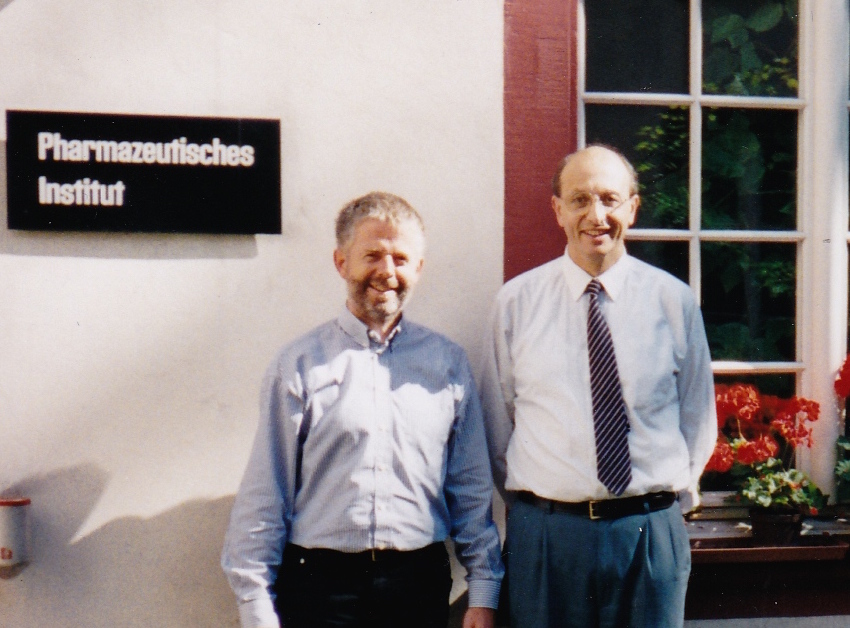
J.R.H. with Alfons Haedener, Basel, 1998.
A big structural research theme developed with the lobster coloration protein crustacyanin; this was work carried out in collaboration with Naomi Chayen at Imperial College and Peter Zagalsky at Royal Holloway College. I also had a new PhD student with me in Manchester, Michele Cianci from Padova University, Italy and also an EU funded research visitor from Poland, Andrzej Olczak. Along with Pierre Rizkallah at Daresbury we solved the crystal structure of apocrustacyanin A1 utilizing softer X rays (λ = 2.0 Å), at SRS PX7.2 (which Pierre had "un moth-balled"), SRS PX9.6 and SRSPX9.5. With the apocrustacyanin A1 crystal structure we solved the beta-crustacyanin crystal structure using data recorded at SRS MPW 14 (this latter beamline development was led by Colin Nave). Our lobster crustacyanin research, published in PNAS in 2002, hit the media! Articles appeared in The Times, The Guardian, The Independent and it was featured also on radio and TV. Later science writers gave their descriptions, e.g., in Physics Today (written by Charles Day), which I especially liked. Madeleine set about crystallizing numerous carotenoids and considerably expanded the available carotenoid x-ray crystal structures and their associated colors. This combination of chemical crystallography along with the biological crystallography proved to be a strength of our lab. Through the 2000s we both presented these results at a variety of conferences having by now been able to travel together, as our children had grown up and "flown the nest".
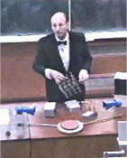
On the occasion of the 150thAnniversary of the University of Manchester, at their invitation here I am in 2001 as their "W. L. Bragg Lecturer". (W. L. Bragg was Professor in the Manchester Physics Department from 1919 to 1937.)
The x-ray laser arrival was clearly very exciting, I thought, from the outset. In 2002 I was Director of SR Science based full time at Daresbury. I firmly encouraged UK participation. In 2004 I published some ideas of how one might optimize its use in protein crystallography in a conference proceedings paper (the International Symposium on Diffraction Structural Biology, ISDSB, held in 2003 in Japan) in the Journal of Synchrotron Radiation. My paper describes finding "marker" seleno atoms in Se-met amino acid residues from two wavelength dispersive differences and secondly use of longer wavelengths along with back scattering to still realize at least 2.5 Å diffraction resolution. In general, though, the biological crystallography community reaction to the x-ray laser was as controversial as the earlier reaction on whether synchrotron radiation would be useful in protein crystallography! In Manchester, with my final year undergraduate project and master students, we started investigating electron-rich heavy-atom clusters binding to lysozyme as a test protein; tantalum bromide (Ta6Br12) and the platinum hexahalides, especially platinum hexaiodide, were interesting. My idea was that with protein samples as small as a single molecule in the x-ray laser beam their diffraction patterns would be exceedingly weak and so we would need both recognizable markers, like the Se-met above, and the nicely shaped octahedral platinum hexahalides. These would also provide an increase in the x-ray scattering efficiency of a protein sample. I spoke on these ideas and results at the ECM28 in 2013 held in UK. At the x-ray laser session in Albuquerque I made the additional suggestion that fully loaded ferritin, with its protein shell enclosing some 2000 iron atoms, would make a splendid test "nanocluster" to spray past the Linac Coherent Light Source LCLS Stanford x-ray laser beam. John Spence, who sat nearby, looked at me and said "ferrritin is planned".
J.R.H. at the 2005 IUCr meeting in Florence. I heartily thank all my PhD students and post docs over the last 35 years for the research and development work we have undertaken, as well as the people I mentioned above. I would like to mention that I collaborated for many years with George Habash, Durward Cruickshank and Jim Raftery. I am very grateful to CCP4 whose software I have extensively relied upon, as well as my students and I learning a great deal at their Study Weekends, as well as SHELX software (for full matrix inversions) and Xplor/Phenix software (for neutron macromolecular crystallography). I also heartily thank Daresbury Laboratory and the universities of York, Oxford, Keele and Manchester; and all the SR and neutron facilities for their collaboration since 1976. I especially thank my wife Madeleine.
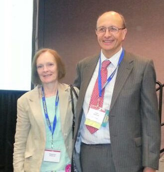
Madeleine and J.R.H. at the 2014 ACA meeting in Albuquerque. When I received the email from the ACA President Cheryl Stevens that I had been selected to receive the 2014 ACA Patterson Award, I immediately told Madeleine that I could not have achieved it without her. The ACA conference in Albuquerque was, of course, a wonderful experience. As well as the Patterson Award Lecture I gave two other lectures, on the use of high-photon energies in macromolecular crystallography and on involving undergraduates in protein crystallography research. My past PhDs (Dora Gomez, Michele Cianci, and Eddie Snell) and collaborators (Zygmunt and Ulla Derewenda, Howard Einspahr and Keith Moffat) hosted a marvelous dinner for Madeleine and me near the Albquerque Convention Center.. I was proud also to enter the ACA 2014 Conference Banquet alongside the ACA President Prof Martha Teeter. As with all the ACA meetings I have attended over the years, I greatly enjoyed all of this ACA meeting. Thank you to the ACA!
[1] Hodgkin, Dorothy. In Patterson and Pattersons: Fifty Years of the Patterson Function; Glusker, Jenny P., Patterson, Betty K., Rossi, Miriam, Eds.; Oxford: New York, 1987; p 637.
[2] Phillips, J. C., Wlodawer, A., Yevitz, M. M. and Hodgson, K. O. (1976) PNAS, USA 73, 128-132.
[3] Rosenbaum, G., Holmes, K.C. and Witz, J. (1971) Nature 230, 434-437. |